(1041-B) The influence of TNF-α on Natural Killer cells and the differences between single cells
Thursday, May 25, 2023
13:30 - 14:30 CET
Location: Hall 3
Abstract: NK Cells: Marathon Runners and Couch Potatoes
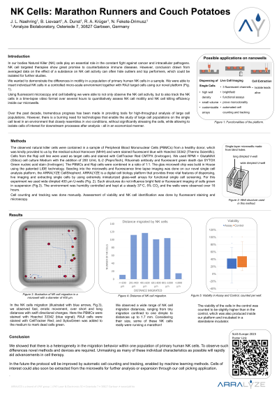
In our bodies Natural Killer (NK) cells play an essential role in the constant fight against cancer and intracellular pathogens. Their potential to eliminate other malignant cells selectively and efficiently is modulated by a complex network of factors and other immune cells. NK cell targeted therapies show great promise to counterbalance immune diseases. However, conclusion drawn from averaged data on the effect of a substance on NK cell activity can often hide outliers and top performers, which could be isolated for further studies. We wanted to demonstrate the differences in motility in a population of primary human NK cells in a sample. We were able to insert individual NK cells in a controlled micro-scale environment together with RAJI target cell.. With our novel platform using inert glass nano-wells, which provide ideal optical properties for microscopy and observation, we were able to insert individual NK cells in a controlled micro-scale environment together with RAJI target cells. Using fluorescent microscopy and cell labelling we were able to not only observe the NK cell activity, but to also track the NK cells using machine learning algorithms in a time-lapse video format over several hours to quantitatively assess NK cell activity and killing efficacy. This way we studied the effect of time in culture and different TNF-α concentrations on primary NK cell activity and the differences between individual NK cells taken from one sample of primary PBMCs. Our experiments showed vast differences in NK cell migration distance in one sample of PBMCs , with some immune cells barely moving and other NK cells becoming true marathon runners with impressive travel distances. With our poster we will demonstrate our findings in cell activity and killing efficacy of individual NK cells in a sample of PBMCs with imposing fluorescent microscopy images with cell tracking overlay and graphic representation of our data. We will also show how we used the micrometer-sized glass nano-wells to conduct our analysis and acquire high quality microscopy images for the tracking algorithm. Our contribution will be in particular of interest to participants in the fields of immunology, therapeutic antibodies, cell therapy, cell line development and SynBio.